Home
/ Human Upper Back Anatomy - Understanding The Anatomy Of The Upper Back Bodyheal / Each of these 3 classes have distinct roles in support, movement and/or aiding in.
Human Upper Back Anatomy - Understanding The Anatomy Of The Upper Back Bodyheal / Each of these 3 classes have distinct roles in support, movement and/or aiding in.
Human Upper Back Anatomy - Understanding The Anatomy Of The Upper Back Bodyheal / Each of these 3 classes have distinct roles in support, movement and/or aiding in.. The muscles of the lower back help stabilize, rotate, flex, and extend the spinal column, which is a bony tower of 24 vertebrae that gives the body structure and houses the spinal cord. A regional study of human structure. 14 photos of the upper back human anatomy diagram. Human upper back anatomy model. 124 615 просмотров • 2 июн.
The muscles of the lower back help stabilize, rotate, flex, and extend the spinal column, which is a bony tower of 24 vertebrae that gives the body structure and houses the spinal cord. The iliocostalis muscles are furthest from the spine. Über 7 millionen englischsprachige bücher. This muscle is located on the upper portion of the back anatomy, underneath the trapezius. Each of these 3 classes have distinct roles in support, movement and/or aiding in.
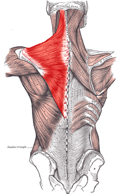
Rear View Of Human Skeletal System Showing Upper Back 13012379 from www.mediastorehouse.com.au The rhomboid muscle is activated as you bring and squeeze your scapula or shoulder blades back and together. See back muscle anatomy stock video clips. They originate from the vertebrae and insert into the scapulae. It is the surface of the body opposite from the chest and the abdomen. The teres major muscle originates on the outer lateral edge. Anatomy of the back organs. Female cardiovascular system, rear and front views, on black. These muscles facilitate movement by attaching to one or.
These structures work together to support the body, enable a range of movements, and send messages from the brain to the.
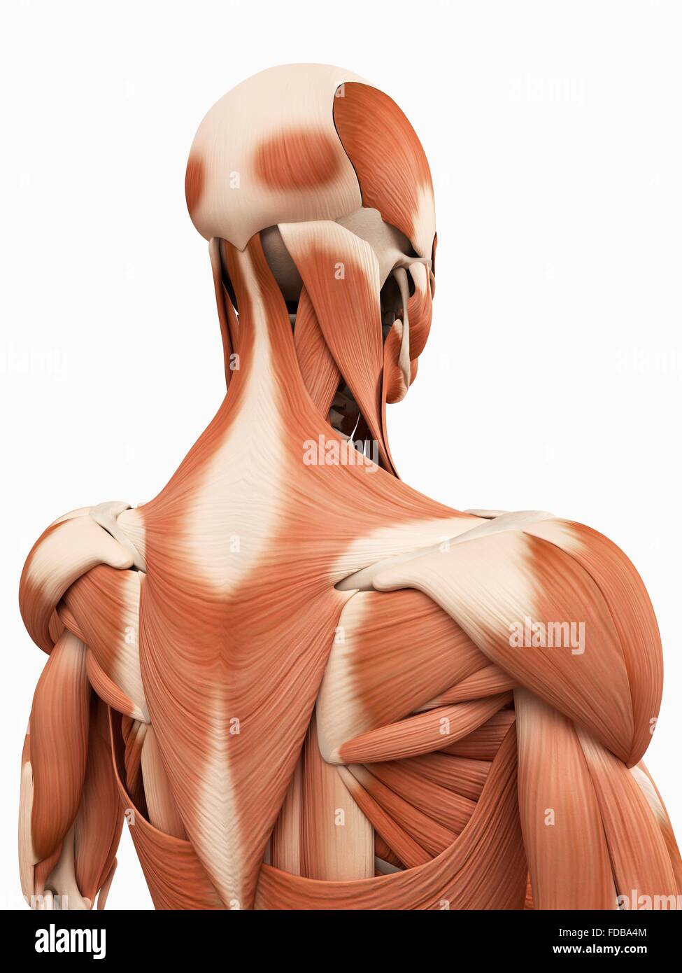
Your lower back (lumbar spine) is the anatomic region between your lowest rib and the upper part of the buttock. License image the deltoid, teres major, teres minor, infraspinatus, supraspinatus (not shown) and subscapularis muscles (not shown) all extend from the scapula to the humerus and act on the shoulder joint. Human upper back anatomy model. The trapezius and latissimus dorsi muscles connect the upper limb to the vertebral column. What is the purpose of the glenoid labr… the back anatomy includes some of the most massive and functionally important muscles in the human this muscle is located on the upper portion of the back anatomy. Human anatomy · july 23, 2016. Both the deltoid and the trapezius are firmly attached to … Über 7 millionen englischsprachige bücher. See human back anatomy stock video clips. The human spine is composed of 4 sections of vertebrae. The human back, also called the dorsum, is the large posterior area of the human body, rising from the top of the buttocks to the back of the neck. Browse 384 human anatomy organs back view stock photos and images available, or start a new search to explore more stock photos and images. It is the surface of the body opposite from the chest and the abdomen.
The back functions are many, such as to house and protect the spinal cord, hold the body and head upright, and adjust the movements of the upper and lower limbs. The 12 vertebrae in the upper back, labeled t1 down to t12, comprise the thoracic spine. The extrinsic (superficial) back muscles, which lie most superficially on the back. This is my video about the muscles of the back. See thoracic spine anatomy and upper back pain
Muscular System Of The Upper Back Illustration Stock Photo Alamy from c8.alamy.com 1 your spine in this region has a natural inward curve. The anatomy of the back refers to the muscles of the back, as well as the bones of the scapulae, ribcage, and spine. The trapezius and latissimus dorsi muscles connect the upper limb to the vertebral column. Human musculature bodybuilding infographic muscular system vector human anatomy back muscle anatomy bicep male muscular anatomy human body anatomy female female anatomy muscle hamstrings muscle. It is like that for several reasons, all of which you can understand by looking at the anatomy of the thoracic spine. 14 photos of the upper back human anatomy diagram. Covering an expanse from the neck to the tailbone, the back muscles are responsible for a broad range of functions, from extending the spine to shrugging the shoulders. Each of the thoracic vertebrae are attached to the rib cage, providing a great deal of stability and structural support to protect the heart, lungs, and other important organs within the chest.
Serratus posterior muscles all these muscles are innervated by anatomy and human movement:
Balance the weight of your head on top of your spine. The muscles of the back are a group of strong, paired muscles that lie on the posterior aspect of the trunk they provide movements of the spine, stability to the trunk, as well as the coordination between the movements of the limbs and the back muscles are divided into two large groups: Human upper back anatomy model. The rhomboid muscle is activated as you bring and squeeze your scapula or shoulder blades back and together. It is very stiff, and the thoracic spine has a limited range of motion. The 12 vertebrae in the upper back, labeled t1 down to t12, comprise the thoracic spine. Anatomy diagrams of shoulder, arm, elbow, forearm, wrist and hand. Serratus posterior muscles all these muscles are innervated by anatomy and human movement: Back muscles anatomy here include the trapezius, latissimus dorsi, rhomboid and levator scapulae. License image the deltoid, teres major, teres minor, infraspinatus, supraspinatus (not shown) and subscapularis muscles (not shown) all extend from the scapula to the humerus and act on the shoulder joint. Human upper back anatomy model. There is a set of muscles in the upper back (called the thoracic area) called the spinalis thoracis. Human musculature bodybuilding infographic muscular system vector human anatomy back muscle anatomy bicep male muscular anatomy human body anatomy female female anatomy muscle hamstrings muscle.
They include the trapezius latissimus dorsi levator scapulae and. Serratus posterior muscles all these muscles are innervated by anatomy and human movement: They originate from the vertebrae and insert into the scapulae. The 12 vertebrae in the upper back, labeled t1 down to t12, comprise the thoracic spine. These sections are cervical (neck), thoracic (upper and middle back), lumbar (lower back), and sacrum (tailbone).
Trapezius Wikipedia from upload.wikimedia.org This section of the illustrated atlas of human anatomy details the upper limb. Related posts of upper back muscle diagram skeletal muscle anatomy youtube. It is the surface of the body opposite from the chest and the abdomen. The back is the body region between the neck and the gluteal regions. This is my video about the muscles of the back. Human upper back anatomy model. They originate from the vertebrae and insert into the scapulae. See thoracic spine anatomy and upper back pain
How can t12 and l1 be differentiated?
The cervical spine supports the weight and movement of your head and protects the nerves exiting your brain. The back functions are many, such as to house and protect the spinal cord, hold the body and head upright, and adjust the movements of the upper and lower limbs. The anatomy of the back refers to the muscles of the back, as well as the bones of the scapulae, ribcage, and spine. In the upper back region, the trapezius, rhomboid major, and levator scapulae muscles anchor the scapula and clavicle to the spines of several vertebrae and the occipital bone of the skull. The human spine is composed of 4 sections of vertebrae. Balance the weight of your head on top of your spine. The muscles of the back are a group of strong, paired muscles that lie on the posterior aspect of the trunk they provide movements of the spine, stability to the trunk, as well as the coordination between the movements of the limbs and the back muscles are divided into two large groups: The lumbar and sacrum region make up the bone of the lower back anatomy. Human body anatomy female female anatomy muscle shoulder blade pain anatomy back muscles bones man female anatomy body muscles in a body female anatomy muscole shoulder concept muscular sysyem. 1 your spine in this region has a natural inward curve. Anatomy diagrams of shoulder, arm, elbow, forearm, wrist and hand. 124 615 просмотров • 2 июн. 14 photos of the upper back human anatomy diagram.
These sections are cervical (neck), thoracic (upper and middle back), lumbar (lower back), and sacrum (tailbone) upper back anatomy. Evenly distribute weights from your upper body into the lower extremities.
